INCHAE
/

코인두

비인두
라틴어Pars nasalis pharyngis영어NasopharynxTA22862
3D로 보기

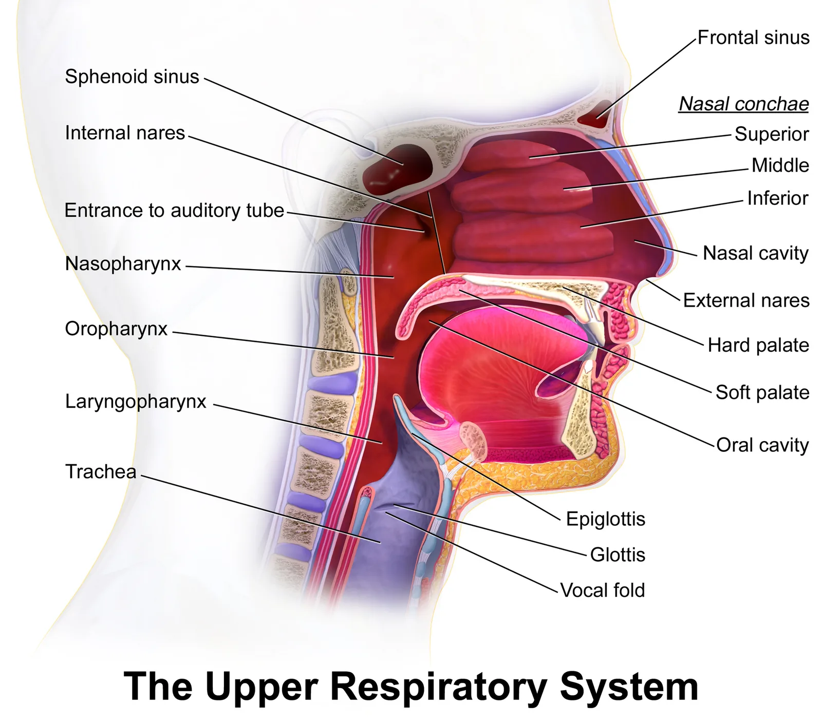
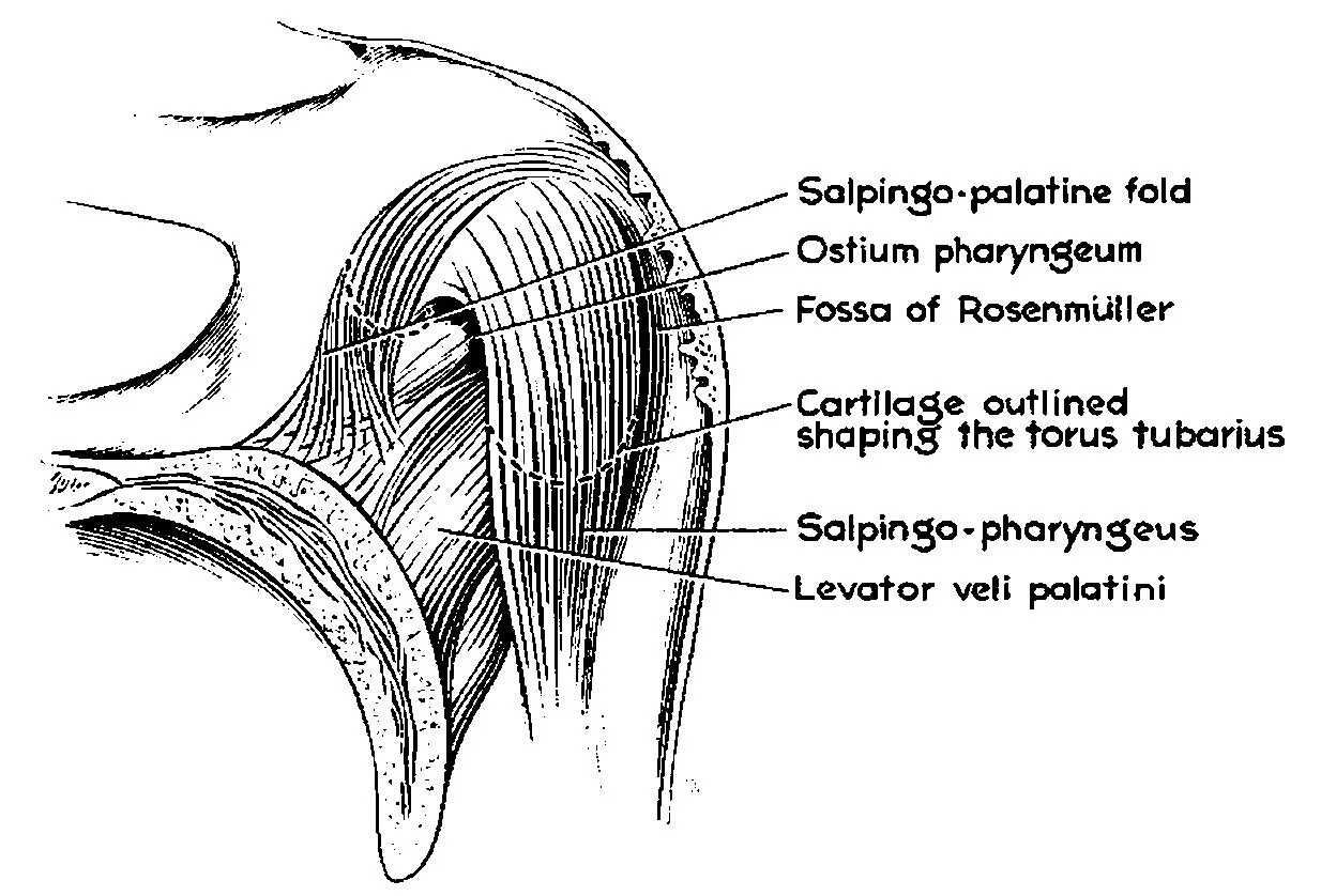
뒤콧구멍 뒤에서 물렁입천장까지의 인두 윗부분으로, 호흡만 하는 길이다. 옆벽에 귀관인두구멍과 그 뒤의 귀관융기·인두오목(Rosenmüller 오목), 뒤위벽에 인두편도(아데노이드)가 있다.

핵심 정보

경계
위 = 나비뼈몸통·뒤통수뼈 바닥, 아래 = 물렁입천장(코인두협부), 앞 = 뒤콧구멍
신경
감각 = 위턱신경 인두가지(V2)
림프
인두뒤림프절 → 위깊은목림프절
임상
코인두암은 인두오목에서 잘 생겨 목림프절 비대·한쪽 중이 삼출로 처음 나타난다. 아데노이드 비대는 코막힘·중이염

그림

도해: 위호흡계통 — 가운데귀 근처 귀관 입구
도해: 위호흡계통 — 가운데귀 근처 귀관 입구BruceBlaus . When using this image in external sources it can be cited as: Blausen.com staff (2014). " Medical gallery of Blausen Medical 2014 ". WikiJournal of · CC BY 3.0 · 출처
사진: 귀관융기 해부 세부
사진: 귀관융기 해부 세부S. SIMKINS, PH.D. · Public domain · 출처
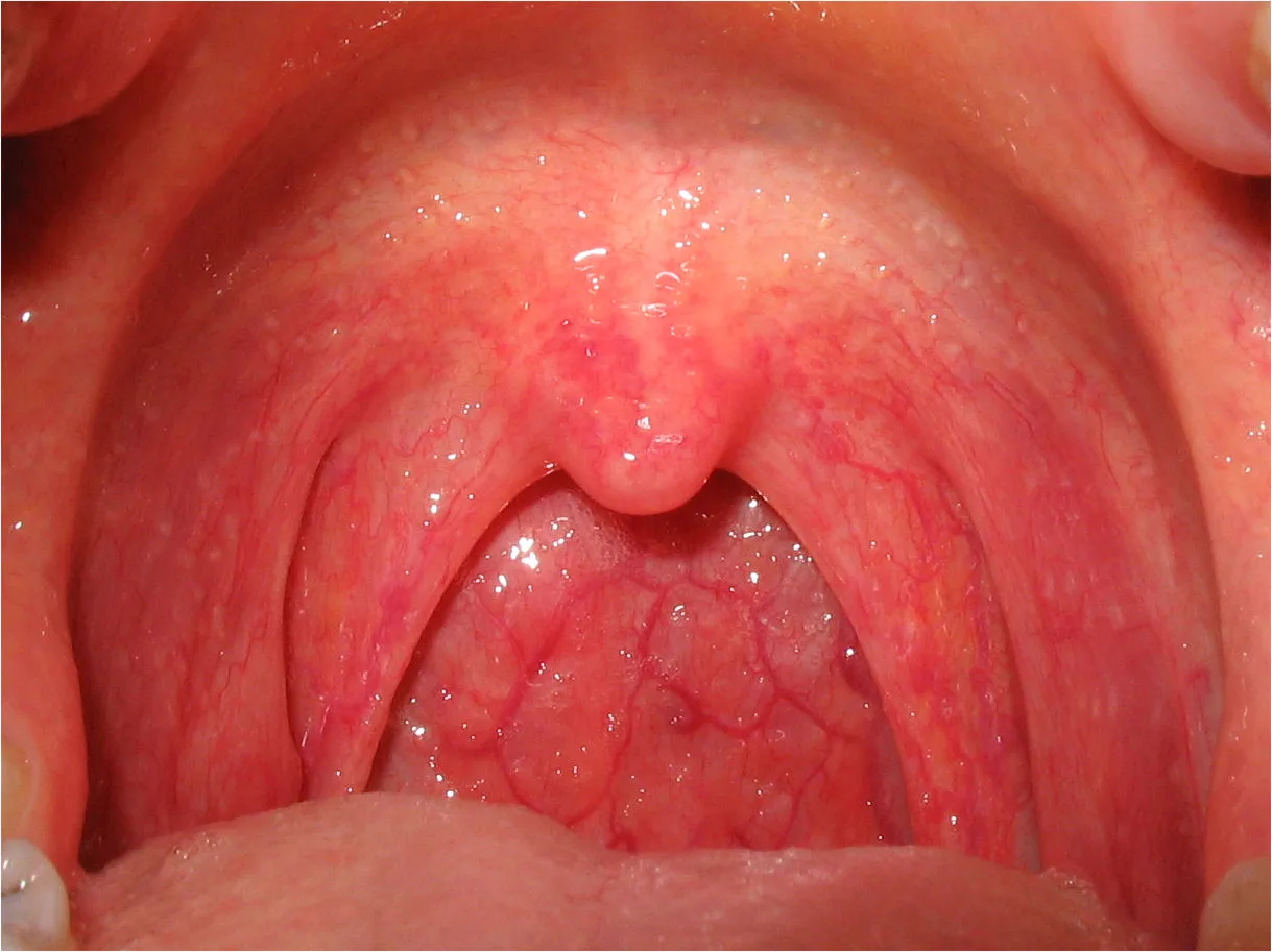
사진: 인두염 — 염증으로 붉게 부은 입인두
사진: 인두염 — 염증으로 붉게 부은 입인두 · CC BY-SA 2.5 · 출처
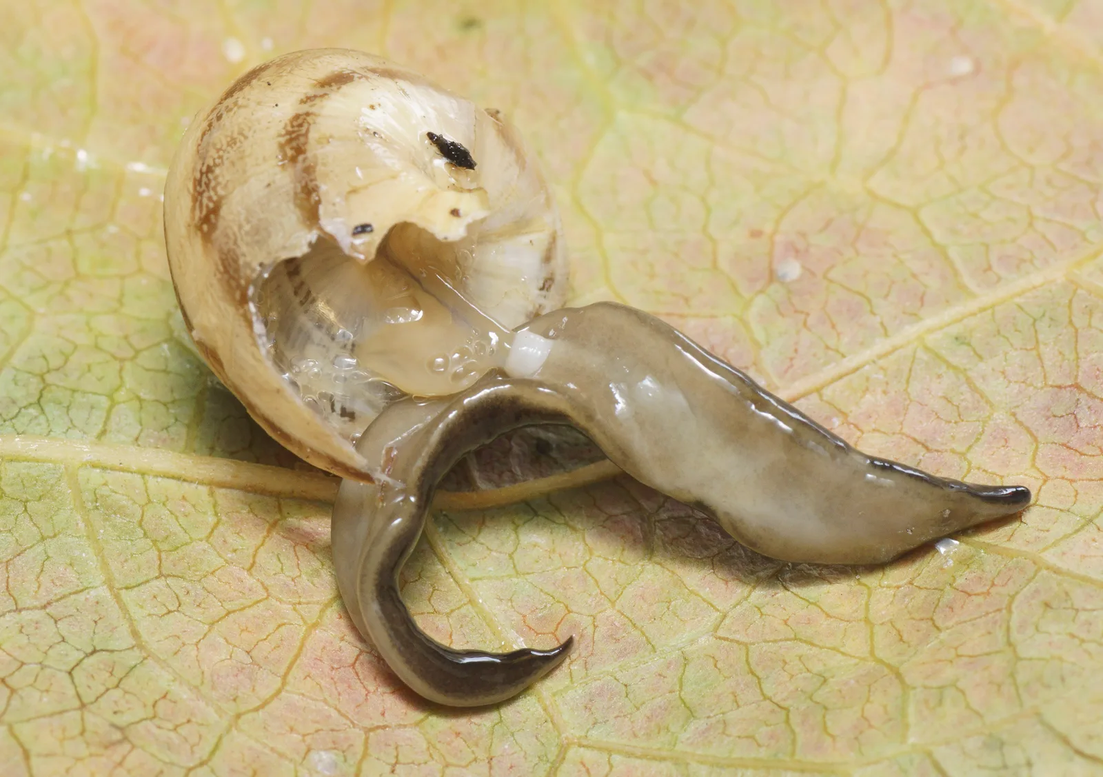
Platydemus manokwari de Beauchamp, 1963, experimental predation on indigenous snail. The flatworm is preying on a snail: it has been disturbed, thus showing the white cylindrical pharynx on the ventral side, protruding and ingesting soft tissues of the snail. The prey is the helicid Eobania vermicul
Platydemus manokwari de Beauchamp, 1963, experimental predation on indigenous snail. The flatworm is preying on a snail: it has been disturbed, thus showing the white cylindrical pharynx on the ventral side, protruding and ingesting soft tissues of the snail. The prey is the helicid Eobania vermiculPierre Gros · CC BY 4.0 · 출처
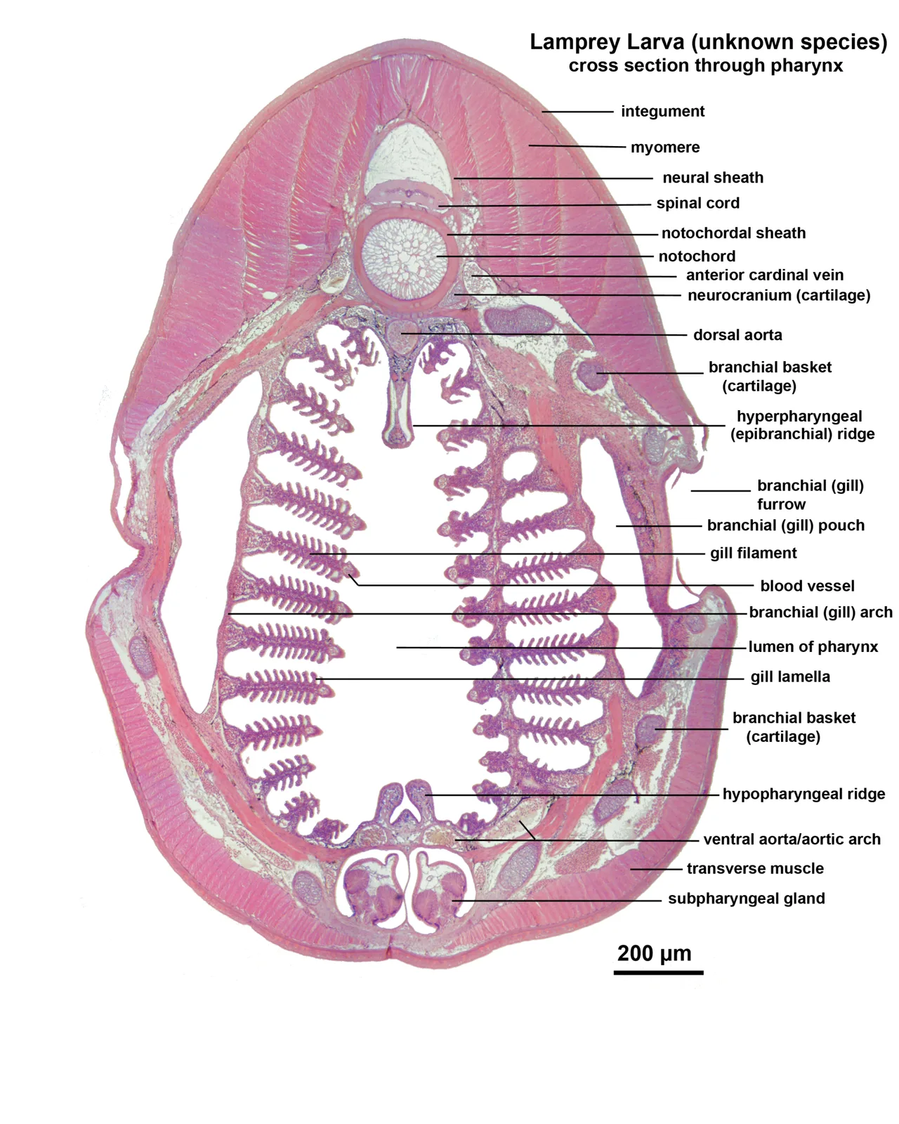
Microscopic cross section through the pharynx of a larva from an unknown lamprey species.
Microscopic cross section through the pharynx of a larva from an unknown lamprey species.Tracyanne · CC BY-SA 3.0 · 출처
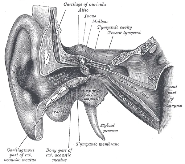
도해: 귀 구조의 단면
도해: 귀 구조의 단면Henry Vandyke Carter · Public domain · 출처

참고 영상 3

유튜브 영상은 각 채널의 것입니다. 구독자 10만 이상 의학 교육·대학병원·공영방송 채널에서 골랐습니다.

참고 문헌

  • Gray's Anatomy 42판
  • Moore 임상해부학 9판